
Your Family’s Eye Care & Eyewear Destination
We’re proud to be a community-centered practice serving families in Galax and the surrounding region. Our compassionate, knowledgeable team is dedicated to providing the most up to date comprehensive eye care. Whether you’re looking for stylish specs or eye disease management we offer a full scope of services!


Focused on Preserving Your Vision
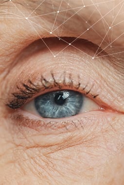
Good vision is more than clear eyesight. We’re here to help safeguard your eye health, preserving comfortable and healthy vision. It all begins with personalization. We listen to your needs and use diagnostic technology to evaluate your eye health thoroughly.
When you visit us, it’s all about what you and your family need. We’re here to support and educate. After all, it’s your vision, and you deserve to be involved in every decision.
Our Location
Visit Us
Our building is located at the intersection of East Stuart Drive and Larkspur Lane. We look forward to welcoming you to our practice family.
Our Address
- 800 E Stuart Dr.
- Galax, VA 24333
Contact Information
- Phone: 276-236-4171
Our Hours
- Monday: 8:00 AM – 6:00 PM
- Tuesday: 8:00 AM – 6:00 PM
- Wednesday: 8:00 AM – 6:00 PM
- Thursday: 8:00 AM – 6:00 PM
- Friday: 8:00 AM – 6:00 PM
- Saturday: Closed
- Sunday: Closed


Our Brands


Our Google Reviews
-
Blue Ridge Eye Care
- 800 E Stuart Dr.
- Galax, VA 24333
-
P: 276-236-4171